In the ever-evolving world of dentistry, technological advancements are making it easier for dentists to provide accurate diagnoses and offer enhanced patient care. One of the most significant improvements in recent years is the advent of digital x-ray technology. Traditional X-rays have been a cornerstone of dental diagnostics for decades, but digital X-rays are revolutionizing the field with improved clarity, safety, and efficiency. At Dickey Dental in Rockhill, SC, Dr. Hugh Brad Dickey utilizes state-of-the-art digital X-ray technology to enhance the dental experience for patients, ensuring accurate diagnostics and improved treatment outcomes.
The Basics: What Are Digital X-Rays?
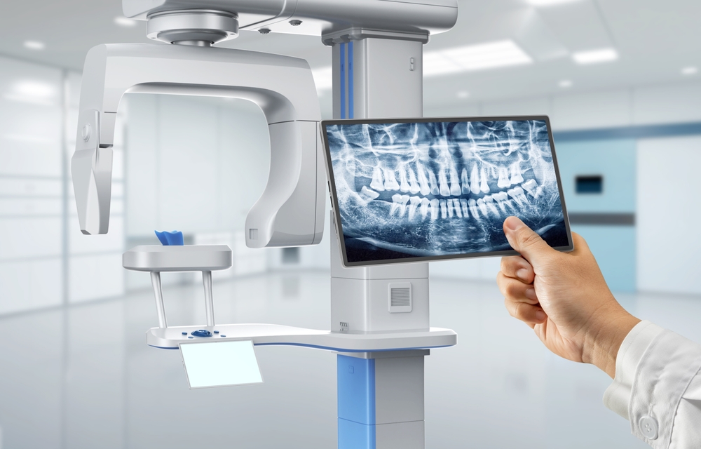
Digital X-rays use electronic sensors rather than traditional film to capture images of the teeth, bones, and surrounding structures. These images are instantly available on a computer screen, allowing Dr. Hugh Brad Dickey to view, enlarge, and manipulate them for better clarity and accuracy.
Unlike traditional X-rays, which use film that must be developed chemically, digital X-rays provide several advantages in terms of speed, safety, and diagnostic capability. As a result, digital imaging has become the preferred method for dentists and patients, revolutionizing how oral health issues are detected and treated.
Benefits of Digital X-Rays
The advantages of digital X-rays extend beyond just convenience. Here are some key benefits of this technology and why it matters for your dental care at Dickey Dental:
- Lower Radiation Exposure One of the most significant advantages of digital X-rays over traditional ones is that they reduce radiation exposure by up to 90%. While dental X-rays have always used low doses of radiation, digital X-rays make the process even safer for patients. This is especially important for children and those who may require multiple X-rays over time.
- With digital X-rays, there is no need to wait for film to develop. The images appear instantly on the dentist’s computer screen, allowing for faster diagnostics. This means that any issues can be identified and discussed in real-time, leading to quicker treatment decisions and minimizing patients’ time in the dental chair.
- Enhanced Image Quality Digital X-rays offer superior image quality, providing Dr. Hugh Brad Dickey with highly detailed and precise images of your teeth and gums. These images can be zoomed in or enhanced to identify the smallest areas of concern, such as tiny cavities, hairline fractures, or hidden infections that might be missed with traditional X-rays.
- Environmentally Friendly Traditional X-rays require the use of chemicals to process the film, which can be harmful to the environment. Digital X-rays eliminate the need for film and chemicals, reducing the environmental footprint of dental offices like Dickey Dental. Additionally, there is no need for physical storage space for X-ray films, as digital images can be stored securely in a computer system.
- Improved Diagnostic Capabilities Digital X-rays allow for advanced diagnostic tools that can aid in detecting oral health issues. For example, digital images can be adjusted to highlight certain areas, making it easier to spot potential problems that may have otherwise gone unnoticed. Dentists can also use software that provides enhanced measurements and comparisons over time, improving the overall accuracy of their diagnoses.
- Easier Sharing and Collaboration Digital X-rays make it simple for dentists to share images with other dental or medical professionals if needed. This is particularly helpful if a patient requires specialized care or a second opinion. At Dickey Dental, Dr. Dickey can easily send images to specialists or include them in electronic medical records, ensuring seamless communication and coordinated care.
Types of Digital X-Rays
Digital X-ray technology encompasses a variety of imaging techniques, each offering unique advantages depending on the situation. At Dickey Dental, Dr. Hugh Brad Dickey uses a combination of these X-ray types to provide comprehensive diagnostics for his patients:
- Intraoral X-rays Intraoral X-rays are the most common type of dental X-ray. These images are taken inside the mouth and provide detailed views of individual teeth, roots, and the surrounding bone. They are excellent for detecting cavities, monitoring the health of the root and bone, and assessing the development of teeth.
- Extraoral X-rays Unlike intraoral X-rays, extraoral X-rays are taken outside the mouth and are used to capture larger areas of the jaw and skull. These X-rays are helpful for evaluating jaw alignment, monitoring impacted teeth, and diagnosing TMJ disorders. While they do not provide as much detail about individual teeth, they offer a broader view of the overall oral structure.
- Panoramic X-rays Panoramic X-rays take a single image of the entire mouth, including the teeth, jaw, and surrounding tissues. This type of x-ray is valuable for identifying impacted teeth, assessing bone loss due to gum disease, and evaluating the need for dental implants. Panoramic images can also help detect oral tumors or cysts, making them an important tool for comprehensive dental exams.
- Cephalometric X-rays Cephalometric X-rays are specialized images used primarily in orthodontics. They show a side view of the skull and are helpful for planning orthodontic treatments, such as braces or corrective jaw surgery. They also provide valuable insights into the alignment and growth of the jaw in relation to the teeth.
How Digital X-Rays Improve Patient Care at Dickey Dental
At Dickey Dental in Rockhill, SC, Dr. Hugh Brad Dickey uses digital X-ray technology to provide patients with top-notch dental care. By embracing the latest advancements in digital imaging, he can offer more accurate diagnoses and personalized treatment plans.
One key benefit of digital X-rays at Dickey Dental is the enhanced ability to detect problems in their earliest stages. Early detection of issues such as tooth decay, gum disease, and bone loss allows for less invasive treatments and a higher likelihood of successful outcomes. Patients also benefit from the lower radiation exposure, making routine X-rays safer over the long term.
Additionally, the instant availability of images enables Dr. Dickey to walk patients through their results immediately. Patients can see what the dentist sees, allowing them to better understand their oral health and make informed decisions about their treatment options.
The Future of Digital X-Ray Technology
Digital X-ray technology constantly advances, and the future holds even more exciting possibilities for improved patient care. Developments in 3D imaging, for example, are already transforming how dentists visualize complex structures like nerves, sinuses, and bone density. These advancements will allow for even more precise treatment planning, particularly in implant dentistry and orthodontics.
Furthermore, as artificial intelligence (AI) becomes more integrated into dental technology, digital X-rays will likely play a key role in helping dentists diagnose and predict oral health issues more accurately. AI algorithms can analyze X-ray images to detect patterns that may not be immediately visible to the human eye, further enhancing the diagnostic process.
Embrace the Benefits of Digital X-Rays at Dickey Dental
Digital X-ray technology has transformed the way dental care is delivered, offering faster, safer, and more accurate diagnostics than ever before. At Dickey Dental, Dr. Hugh Brad Dickey is committed to providing patients in Rockhill, SC, with the highest level of care by utilizing this advanced technology. Whether you need a routine exam or more specialized treatment, digital X-rays can be critical in ensuring that your oral health is in the best possible hands.
To learn more about how digital X-rays can benefit your next dental visit, contact our office at Dickey Dental today to schedule an appointment.
Sources:
- Wenzel, A. (2006). Digital radiography and caries diagnosis. Dental Clinics of North America, 50(4), 735-747.
- Berkhout, W. E., Suomalainen, A., & Jacobs, R. (2012). Digital panoramic radiography: A review of the literature. Dentomaxillofacial Radiology, 41(6), 390-399.
- Farman, A. G., & Farman, T. T. (2005). A comparison of 18 different X-ray detectors currently used in dentistry. Oral Surgery, Medicine, Pathology, Oral Radiology, and Endodontology, 99(4), 485-489.